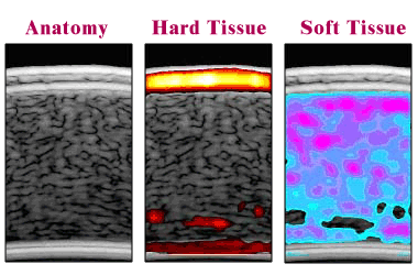
Noninvasive imaging of cornea hardness. An ultrasound-based anatomical image of a small section of the cornea is shown on the left. The center shows hard tissue within the cornea by "electronically staining" those parts of the image measured to be hard. The right shows soft tissue within the cornea by "electronically staining" those parts of the image measured to be soft. Such hardness maps can be used to tailor a LASIK procedure for each individual.