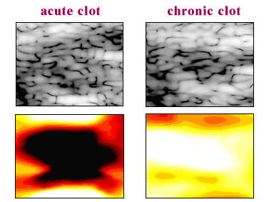
Aging DVT. Top images are conventional ultrasound images of a deep vein showing the presence of a clot. Bottom images are elasticity images, where the large black region in the lower left denotes a soft clot (i.e., young clot) and the bright white region in the lower right denotes a hard clot (i.e., old clot).